Developing Optically-Driven Contractile 3D Human Skeletal Muscle Bioconstruct for an Actuated Joint-on-chip System
PIs:
Dr Ori Bar-Nur, Laboratory of Regenerative and Movement Biology, ETH Zürich
Fellow:
Ali Kerem Kalkan and Prof. Ori Bar-Nur, Laboratory of Regenerative and Movement Biology, ETH Zurich
Collaborators:
Xiaoyu Zhao and Prof. Marcy Zenobi-Wong, Tissue Engineering and Biofabrication, ETH Zurich
Margherita Bernero and Prof. Ralph Müller, Laboratory for Bone Biomechanics, ETH Zurich
Rodrigo Castillo-Acuña and Prof. Dennis Kochmann, Mechanics and Materials Laboratory, ETH Zurich
Goal
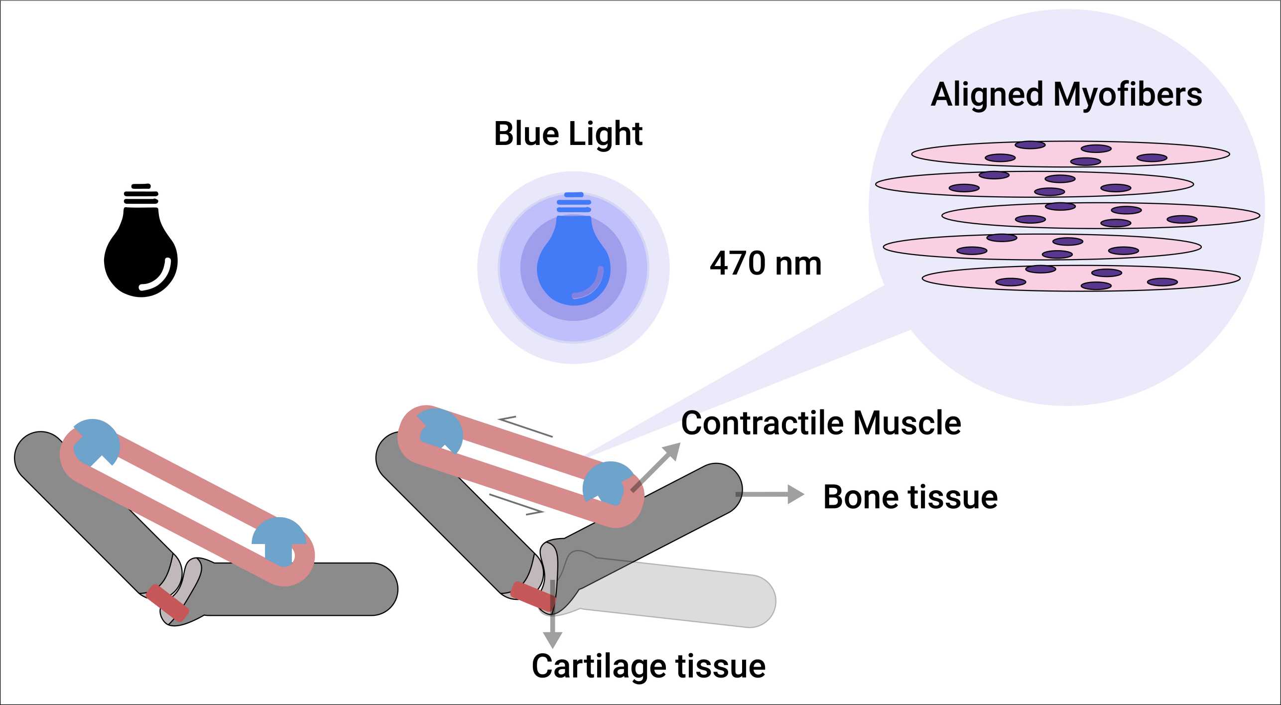
The primary objective of this study is to develop a 3D engineered human muscle bioconstruct integrated with a miniaturized human joint. This construct should accurately replicate the natural process of muscle maturation, encompassing the transformation from myoblasts to mature myofibers, potentially exhibiting regenerative capabilities for tissue repair. Additionally, it should demonstrate the capacity to contract in response to blue light stimulation for a better control of muscle contractility. Our overarching goal is to establish platform that relatively mimics the in vivo conditions related to myofiber alignment, maturation, and contraction. Consequently, this envisioned model holds significant promise for high-throughput applications, thus mitigating the need for animal utilization in medical research.
Methodology
In order to replicate human muscle tissue, our study involves the evaluation of various cell sources, including induced pluripotent cells, fibroblasts, and myoblasts, which have the potential to differentiate into mature muscle cells. To control muscle contraction through optical stimulation, we will employ various delivery techniques to express a light-gated ion channel in myotubes. In order to maximize the force generated by muscle contraction, we will employ 3D printing technologies to fabricate molds suitable for various muscle models that give rise to myotube alignment and maturation. The engineered 3D human muscle bioconstructs will be thoroughly assessed using immunohistochemical staining techniques, allowing us to visualize the cellular components and their organization. Additionally, we will use active force measurement assays to quantitatively evaluate the displacement of the muscle in response to stimulation, thereby assessing its functional capabilities.